Ion channels, the gatekeepers of the cell, are arguably some of the most critical proteins in human biology. Embedded within the fatty membrane of nearly every cell, these intricate protein assemblies regulate the flow of charged ions—such as sodium, potassium, calcium, and chloride—into and out of the cell. This seemingly simple regulatory function is the foundation for virtually all essential physiological processes, including the propagation of electrical signals in the nervous system, the rhythmic contraction of the heart and muscles, the activation of the immune response, and the regulation of cellular volume and pH.
Given their central role in cellular communication and function, it is no surprise that the dysfunction of ion channels is implicated in a vast array of human diseases. These channelopathies span a wide spectrum of disorders, from neurological conditions like epilepsy, pain, and psychiatric disorders, to cardiovascular diseases such as arrhythmias and hypertension, and even specific types of cancer and metabolic disorders. This widespread involvement makes ion channels an exceptionally attractive class of therapeutic targets—in fact, they are the targets of a significant percentage of currently marketed drugs.
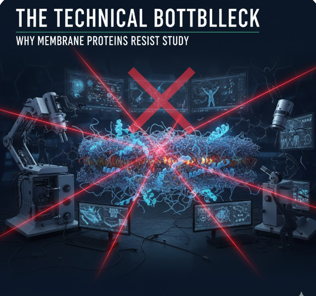
However, the pursuit of new and more effective drugs against these targets has historically been fraught with technical difficulty. Ion channels are membrane proteins, meaning they are naturally situated within the hydrophobic environment of the cell membrane. This inherent location makes them notoriously challenging to study in a stable, biologically relevant state outside of their native environment. Traditional drug discovery methods often require the ion channels to be isolated and purified, a process that can alter their natural structure and behavior, leading to less accurate and often misleading data on how drugs truly interact with them in the body.
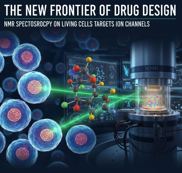
This persistent technical hurdle has been a bottleneck in pharmaceutical development. Now, an international team of researchers, including experts from the University of Seville’s Institute of Chemical Research and collaborators in the UK at the University of East Anglia and the Quadram Institute, has unveiled a game-changing solution. They have pioneered a new technique, based on Nuclear Magnetic Resonance (NMR) spectroscopy, that allows the study of drug-protein interactions directly on living cells, promising to dramatically accelerate the design of potent and highly specific ion channel-targeting drugs.
The Technical Bottleneck: Why Membrane Proteins Resist Study
To understand the magnitude of this new technique, it is essential to first appreciate the challenges posed by membrane proteins in the drug discovery pipeline.
1. Isolation and Instability
The natural environment for an ion channel is the cell membrane, a dynamic lipid bilayer. For traditional structural biology techniques like X-ray crystallography or cryo-electron microscopy (Cryo-EM), the protein must be extracted and solubilized using detergents. This process is complex, costly, and time-consuming. More critically, stripping the protein from its lipid environment can compromise its native three-dimensional structure. A protein’s structure dictates its function, and any alteration can lead to inaccurate binding assays and functional data, meaning a drug that works well on an isolated protein may fail entirely in a living system.
2. Time, Cost, and Complexity
The required preliminary steps—cloning, expression, purification, and stabilization—can take months and require specialized laboratory equipment and highly skilled personnel. This inherent complexity dramatically slows down the structure-activity relationship (SAR) studies, which are the backbone of drug design. SAR studies systematically examine how small chemical changes to a drug molecule (the “key”) affect its therapeutic effect (how well it “opens the lock”). A slow, complex experimental process translates directly to high costs and an elongated drug development timeline.
3. Low Biological Relevance
The most significant limitation of traditional in vitro (outside the living organism) studies is the lack of biological relevance. When a drug is developed, it needs to interact with its target within the complex, crowded, and dynamic environment of a cell membrane. Assays performed on purified proteins miss crucial factors, such as the influence of surrounding membrane lipids, the impact of accessory cellular proteins, and the cell’s own regulatory mechanisms. A drug that looks promising in a test tube might show poor efficacy or unexpected side effects once it encounters the native cellular environment.
The research team, led by experts like Jesús Angulo and Leanne Stokes, recognized that circumventing the need for purification was the key to unlocking faster and more relevant drug design. Their solution harnesses the power of high-resolution NMR spectroscopy.NMR on Living Cells: A New Paradigm for Interaction Analysis
The technique developed by the international research team leverages Nuclear Magnetic Resonance (NMR) spectroscopy, but applies it in a radical new context: directly on the surface of living cells. NMR is a physical phenomenon where nuclei in a magnetic field absorb and re-emit electromagnetic energy. It is a powerful tool for determining the structure of molecules, but traditionally, its application to large, slow-moving molecules like membrane proteins has been challenging.
The researchers adapted a specific NMR method known as Saturation Transfer Difference (STD) NMR spectroscopy. The innovation lies in making this technique work on-cell, effectively transforming a complex biochemical analysis into a simpler, cell-based assay.
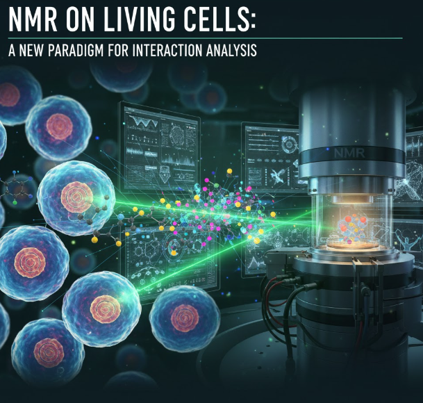
The “On-Cell” Advantage
As summarized by Jesús Angulo of the Institute of Chemical Research, the core advantage is clear: “Until now, studying how drugs interacted with these proteins required isolating them… Our technique, based on nuclear magnetic resonance, allows us to study these interactions in living cells, which provides more biologically relevant information.”
Here is how the new technique provides its revolutionary benefits:
- Elimination of Purification: The most significant time-saver is the complete elimination of the complex and laborious protein purification and isolation steps. The NMR experiments are performed directly on a suspension of living cells, with the ion channels naturally integrated into their cell membranes. This preserves the native environment of the protein, ensuring the drug interactions observed are those that would occur in vivo.
- Speed and Economy: The experiments are remarkably fast, often lasting less than an hour. This speed, combined with the removal of complex pre-processing, makes the technique dramatically more economical and simpler. The researchers anticipate that their method could quickly become a standard, high-throughput tool in pharmacological laboratories globally for rapid structure-activity studies.
- Specific Interaction Mapping: The STD NMR technique provides unprecedented detail. By analyzing the data, researchers can identify exactly which parts of the drug molecule interact with specific binding sites on the cell membrane protein. As Serena Monaco explains, this allows researchers to “optimise these interactions,” which is critical for designing drugs that are not only effective but also highly specific, minimizing off-target effects and potential side effects.
Case Study: Targeting the P2X7 Receptor
To validate the utility of their new technique, the research team applied it to a highly relevant therapeutic target: the P2X7 receptor.
The P2X7 receptor is an adenosine triphosphate (ATP)-gated ion channel that plays a vital role in the immune system, inflammation, and pain transmission. Its dysfunction and overactivity have been strongly linked to a variety of debilitating human conditions, including major depression, certain autism spectrum disorders, chronic inflammatory diseases, and several types of cancer. Due to its broad involvement in disease pathology, the P2X7 receptor is a primary focus for drug development efforts.
Using their on-cell NMR technique, the researchers successfully characterized the binding interactions of a known drug molecule that acts as a negative allosteric modulator (NAM)—a type of drug that binds to a site separate from the ion channel’s main pore to reduce its activity. They confirmed that they could precisely map the binding interface of the NAM drug directly on the P2X7 receptors embedded within the living cell membranes.
This demonstration proved that the technique is robust and sensitive enough to handle the complexity of a natural cellular system, providing high-resolution structural information that was previously only obtainable through tedious, non-native in vitro methods.
Synergy with Bioinformatics: Validating the Digital Drug Key
The research team did not stop at experimental validation; they also integrated their in vivo data with in silico (computer-generated) models, creating a powerful new paradigm for rational drug design.
Modern drug discovery heavily relies on bioinformatics and computational modeling. Researchers use sophisticated software to generate three-dimensional models of drug-receptor binding, essentially creating a library of potential ‘keys’ (drug molecules) and predicting how well they might fit the ‘lock’ (the ion channel).
However, these computer models are only as good as the assumptions they are built upon. An essential step has always been laboratory validation. The team, using software developed at their home institution, combined their experimental on-cell NMR data with the computational binding models. This collaboration allowed them to validate which computer-generated models actually matched the observations made on living cells.
This fusion of experimental and computational data represents a significant leap forward. As Angulo eloquently put it, the process is like a “lock and key” mechanism. “Bioinformatic models are essential to designing new drugs,” he notes, but the ability “to validate three-dimensional computer models on living cells represents a new paradigm in the development of drugs targeting these proteins.” Instead of relying on predictions based on purified, potentially altered protein structures, researchers can now fine-tune their computational designs using highly accurate, biologically relevant data obtained in near real-time. This combination will drastically reduce the failure rate of candidate drugs early in the discovery process.
Future Outlook: Opening New Therapeutic Channels
The development of on-cell STD NMR spectroscopy is more than a technical advancement; it is an enabling technology that promises to open up entirely new research possibilities across multiple disease areas.
1. Expanding the Target Pool: Many challenging membrane proteins beyond ion channels, such as G protein-coupled receptors (GPCRs) and transporters, are also difficult to study using traditional methods. This new NMR technique is highly adaptable and can potentially be applied to accelerate drug discovery against these targets as well, which are collectively involved in nearly 80% of all therapeutic drug actions.
2. Precision Medicine: The ability to rapidly and precisely map drug interactions allows for the design of highly selective compounds. In complex diseases like cancer, where multiple related ion channels might be expressed, the development of a drug that selectively targets only the disease-driving channel will lead to fewer systemic side effects and more effective, patient-specific therapies.

3. Accelerating the Timeline: By eliminating complex purification steps and speeding up the SAR loop, the technology can cut months or even years off the early stages of drug development. This acceleration is critical for bringing new treatments to market faster for devastating diseases like Alzheimer’s, schizophrenia, and heart failure, where dysfunction of ion channels is a known component.
In conclusion, the bottleneck created by the difficulty of studying membrane proteins has long been a major barrier in drug development. This new NMR-based technique, enabling researchers to observe the critical dance between a drug and its ion channel target on the surface of a living cell, represents a significant breakthrough. It is a faster, simpler, and more relevant approach that is set to revolutionize pharmacological studies and usher in a new era of highly effective, highly specific treatments for a host of human diseases.